ICRIRET 2025 · 3rd International Conference on Recent and Innovative Results in Engineering and Technology · Konya
Nucleus Segmentation in Heterogeneous Microscopy Images on the Data Science Bowl 2018 Dataset
Summary
At a Glance
A head-to-head comparison of two real-time instance segmentation models on cell nuclei. The newer, attention-based YOLOv12s-seg loses to the more mature YOLOv11s-seg on accuracy and on speed.
DSB2018: 670 images from bright-field and fluorescence microscopy, 10 to 300+ nuclei each.
Same small variants, 512×512 input, batch 16, 100 epochs, same augmentation and GPU.
Architectural maturity and training stability can outweigh lightweight novelty.
I · Introduction
Why Segment Every Nucleus?
- Nuclei are the basic unit of tissue analysis: counting them and measuring their shape supports cancer diagnosis and biomedical research.
- Manual delineation is slow and varies between experts; automation reduces human error and speeds up diagnostic workflows.
- Instance segmentation is needed, not only semantic: touching nuclei must be separated into individual masks.
- Microscopy is heterogeneous: stains, modalities, magnification and cell types change from image to image.
One mask: "nucleus" vs "background". Touching nuclei become one blob.
One mask per nucleus, so each can be counted and measured. This is what YOLO-seg models produce.
I · Related Work · Interactive
Related Work
Results as reported by each study, on different datasets and metrics, so they are not directly comparable. Click a column header to sort.
| Method | Family | Key idea | Reported result | Ref. |
|---|---|---|---|---|
| BAWGNet | U-Net | wavelet-guided attention, boundary-aware unit | Dice 90.82% (DSB-2018) | 5 |
| ConDANet | U-Net | contourlet-driven attention, wavelet pooling | Dice 88.9 / 81.71 / 75.12% (3 datasets) | 6 |
| ASRDNet | U-Net | dilated separable residual blocks, channel attention | Dice 0.9129 (DSB-2018) | 7 |
| CBAM-Residual U-Net | U-Net | residual convolutions with CBAM attention | above SOTA on DSB-2018, GlaS, TNBC | 8 |
| M-UNet | U-Net | parallel kernel heads, shearlet unsharp masking | IoU 0.8325, F1 0.9086 | 9 |
| U-Net / W-U-Net | U-Net | resizing, normalization, augmentation | Dice 0.9916, IoU 0.9675 | 10 |
| Swin + GCFM | Transformer / graph | graph convolutional feature fusion | above SOTA on DSB-2018, LIVECell | 11 |
| Multi-task network | Transformer / graph | bottleneck transformer, distance maps, stain transfer | better on NuInsSeg, MoNuSeg, DSB-2018 | 12 |
| DPGNet | Transformer / graph | edge difference attention, uncertainty maps | best Boundary-IoU and HD95; 25.51M params | 13 |
| ASF-YOLO | One-stage | scale-sequence fusion, channel-position attention | mask mAP 0.887, 47.3 FPS (DSB-2018) | 14 |
| YOLACT | One-stage | prototype masks + coefficients, no repooling | mAP 29.8 at 33.5 FPS (COCO) | 15 |
| Improved BlendMask | One-stage | dilated and context aggregation, ranking loss | +3.6% AP (DSB-2018) | 16 |
| U-Net++ + watershed | Clinical tool | GUI with interactive refinement and reports | F1 0.917, IoU 0.846 (DSB-2018) | 17 |
| GA-UNet | Clinical tool | genetic algorithm shrinks U-Net to < 1% of parameters | accuracy 95.96% (DSB-2018) | 18 |
Common limits: heavy computation, overfitting on small sets, sensitivity to stain normalization. Gap: no direct YOLOv11-seg vs YOLOv12-seg comparison on nuclei.
II.A · Dataset and Preprocessing
The Data Science Bowl 2018 Dataset
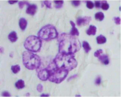
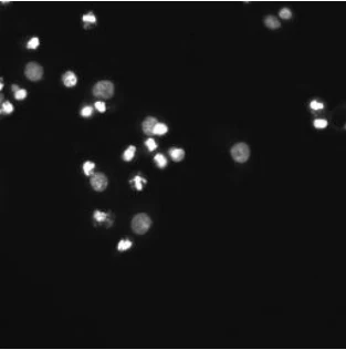
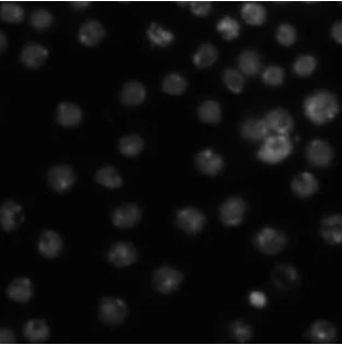
Fig. 1: sample training images. Stains, modality and density change from image to image.
536 training and 134 validation (80/20, stratified by nuclei density).
Bright-field and fluorescence, various stains, magnifications and cell types.
256×256 to 1080×1080 pixels; 10 to more than 300 nuclei per image.
Resized to 512×512 with padding to keep the aspect ratio; one binary mask per nucleus.
II.B–C · Architectures
Mature vs. New: The Two Contenders
YOLOv11s-seg
- CSPNet-based backbone
- Path Aggregation Network (PAN) neck for multi-scale feature fusion
- anchor-free detection head extended with a mask branch
- started from the official COCO-pretrained checkpoint
yolo11s-seg.pt
YOLOv12s-seg
- lightweight, attention-centric backbone with FlashAttention
- redesigned segmentation head for mask refinement
- aims at better feature alignment and lower overhead
- built from
yolo12s-seg.yaml, i.e. from scratch with COCO initialization
II.D · Table 1 · Interactive
Architectural and Computational Efficiency
III.A · Experimental Setup · Table 2
Training Setup
| Parameter | Value |
|---|---|
| Optimizer | AdamW (lr = 0.002) |
| Batch size | 16 |
| Input resolution | 512×512 |
| Epochs | 100, cosine learning-rate schedule |
| Augmentation | mosaic, HSV, flip |
| Precision | automatic mixed precision (AMP) |
Laptop: NVIDIA GeForce RTX 3050 (6 GB VRAM), Intel CPU with 8 cores / 12 threads, 16 GB RAM, CUDA 11.8, PyTorch.
A 6 GB consumer GPU is close to what a small lab or clinic would use; memory and latency differences are felt directly.
III.B · Fig. 2 · Interactive
Confusion Matrices
Counts aggregated over the 134 validation images; the background/background cell is not defined for instance matching. Metrics on this slide are computed from the counts.
III.C · Figs. 3–4 · Interactive
Training Dynamics over 100 Epochs
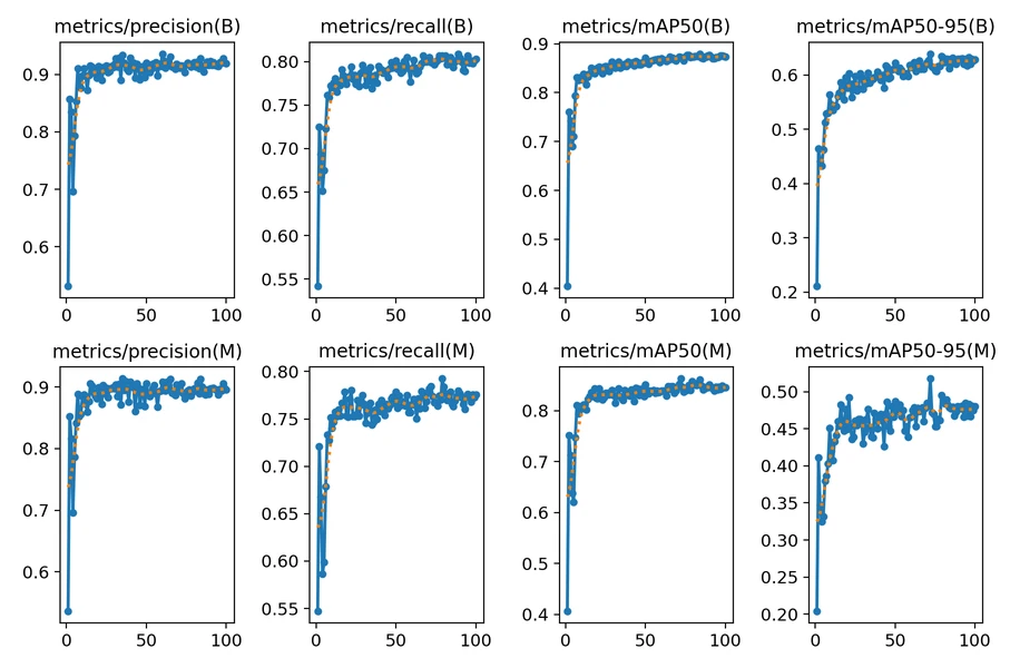
Top row: boxes (B). Bottom row: masks (M).
- Fast gains in the first 20 epochs: COCO pretraining transfers well.
- Plateaus after epoch 60, no sign of overfitting.
- Box curves are nearly identical; the models separate on mask recall and mask mAP@0.5:0.95.
III.D · Metrics · Interactive
IoU Lab: Why mAP@0.5:0.95 Is Strict
A made-up round nucleus, not data from the paper. mAP@0.5:0.95 averages the AP at ten IoU thresholds from 0.50 to 0.95, so it rewards tight boundaries.
III.D–E · Tables 3–4 · Interactive
Validation Metrics
III.E · Fig. 5 · Interactive
Segmentation Results on Validation Images
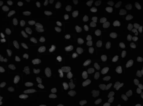
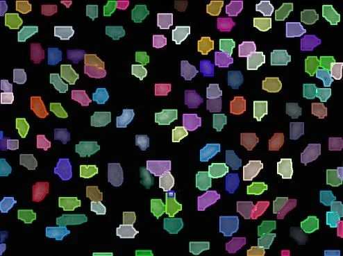
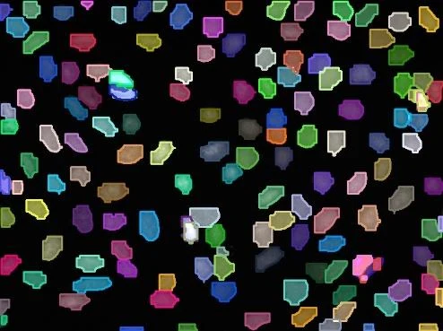
Across the samples, YOLOv11s-seg keeps tight boundaries and separates instances; YOLOv12s-seg shows boundary artifacts, missed small nuclei and occasional merging of adjacent nuclei.
IV · Discussion
Why Does the Older Model Win?
The PAN neck aligns multi-scale features well, which matters for nuclei that range from a few pixels to large blobs.
A tuned, pretrained checkpoint against a YAML build with COCO initialization: YOLOv11s-seg converges earlier (epoch 88 vs 98).
FlashAttention and 91 more layers bring no gain here, yet cost memory (5.52 vs 4.62 GB) and latency (11.3 vs 6.3 ms).
The gap is small at IoU 0.5 and grows at strict thresholds: mask quality, not detection, separates the models.
IV · Conclusion and Future Work
Conclusion
- On heterogeneous DSB2018, YOLOv11s-seg beats YOLOv12s-seg: mask mAP@0.5:0.95 0.528 vs 0.478, mask recall 0.784 vs 0.755, latency 6.3 vs 11.3 ms.
- It is the preferred model for real-time nucleus segmentation under diverse staining and imaging conditions.
Future work
YOLOv11-seg combined with domain-specific attention mechanisms.
Cross-dataset tests on multi-organ histopathology cohorts.
Interactive refinement interfaces for clinical adoption.
IV · Discussion
Check Questions
- Why is instance segmentation, not semantic segmentation, needed for nuclei?
- YOLOv12s-seg has fewer parameters and the same GFLOPs. Why is it still slower?
- From the confusion matrices, which model misses more nuclei, and which produces more false nuclei?
- Why do the models differ more on mAP@0.5:0.95 than on mAP@0.5?
- Name two reasons the paper gives for the advantage of YOLOv11s-seg.
Show answers
- Touching nuclei must become separate objects so that each can be counted and measured.
- It is deeper (294 vs 203 layers) and uses attention; latency depends on depth and memory traffic, not only on FLOPs.
- YOLOv12s-seg misses more (1042 vs 834 false negatives); YOLOv11s-seg produces more false nuclei (1009 vs 855).
- The strict metric also rewards boundary accuracy; small mask errors pass at IoU 0.5 but fail at higher thresholds.
- The mature PAN neck for multi-scale fusion, and stable transfer from a pretrained COCO checkpoint.
References
Selected References
- Goodman, A. et al. (2018). 2018 Data Science Bowl. Kaggle.
- Jocher, G., Qiu, J. (2024). Ultralytics YOLO11. github.com/ultralytics/ultralytics.
- Tian, Y., Ye, Q., Doermann, D. (2025). YOLOv12: Attention-centric real-time object detectors. arXiv:2502.12524.
- Kang, M. et al. (2024). ASF-YOLO: A novel YOLO model with attentional scale sequence fusion for cell instance segmentation. Image Vis. Comput., 147, 105057.
- Imtiaz, T., Fattah, S. A., Kung, S.-Y. (2023). BAWGNet: Boundary aware wavelet guided network for the nuclei segmentation in histopathology images. Comput. Biol. Med., 165, 107378.
- Bolya, D. et al. (2019). YOLACT: Real-time instance segmentation. ICCV, 9156–9165.
See the full paper for the complete reference list. Figures on these slides are from the paper.